The Immune System of the Brain
Though the brain is a powerful computational tool, it is also a sensitive and delicate organ. For this reason, it requires specialized protection. This includes the skull, the membranes covering the brain and spinal cord, and the blood-brain barrier (BBB), which is a specialized system of cells that line the blood vessels of the brain and serve as a filter, allowing useful molecules to enter while keeping harmful things out.
The blood-brain barrier isn’t perfect. Sometimes harmful things can make their way into the brain. In other parts of the body, this problem is managed by the immune system, which circulates through the blood and enters infected areas to kill pathogens and spur healing. However, in the same way that the BBB keeps pathogens out of the brain, it also prevents the body’s immune cells from entering the brain.
So, what happens when something harmful crosses the brain?
Worry not, because the microglia are the brain’s first line of defense.
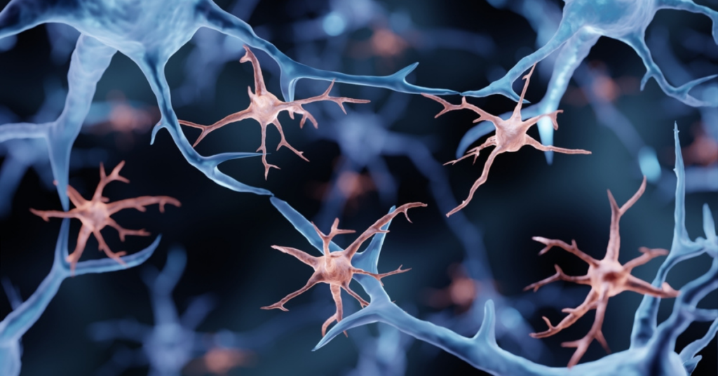
Microglia are immune cells that live only in the brain. They are evenly distributed throughout the brain and spinal cord, making up around 10% of the total cells. Under healthy conditions, microglia have long branches that are used for moving and surveying the surrounding area for pathogens. Even when “resting” they are constantly on the lookout for problems.
The Guardians of the Brain
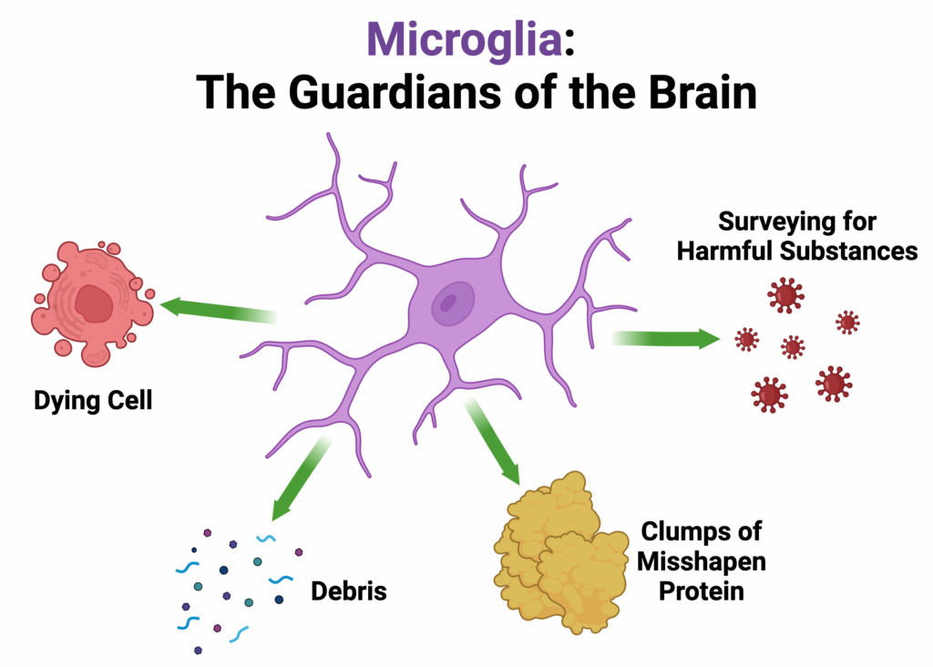
Microglia play many roles in maintaining our brains in a healthy state, such as removing dead or dying brain cells, debris, and clumps of misshapen protein. When microglia detect a nearby injury or threat in their environment, they “activate” to deal with the problem.
Activated microglia are visibly distinct from “resting” microglia by the thickening and shortening of their branches. They release specific inflammatory chemicals that alert other nearby cells to the presence of a problem and serve as a “come help!” signal to other microglia.
Thus, when looking at a brain area where injury has occurred, researchers can often see not only a change in the shape of the microglia but also an increase in the number of microglia that are present. Once the problem is identified (whether a harmful substance or a damaged brain cell), microglia can eliminate the threat by eating, or “phagocytosing”, the materials causing the threat.

An overview of what Microglia do in the brain. Figure made by : Rana Abdelhalim using BioRender.
Microglia in Aging
As we age, changes in the brain take place that may cause a gradual loss of normal functioning. For instance, in the brains of aged individuals, “aged” microglia were observed to have altered morphology, reduced ability to remove debris, dead cells, and misshapen proteins.
They were also observed to have deficits in their surveillance actions to detect harmful substances in the surrounding environment. And when a harmful substance is detected, aged microglia are less likely to move to where the harmful substance is to deal with the problem.
When activated, these aged microglia may also release different types of inflammatory chemicals that heighten inflammation, leading to a negative impact on surrounding cells. These changes in aged microglia are of great interest since they may contribute to the development of neurodegenerative diseases.
Role of Microglia in Ataxias
Ataxias are a class of neurodegenerative diseases characterized by deficits in motor coordination. This is caused by the dysfunction and death of Purkinje cells, which are specialized neurons that drive the function of the cerebellum. As these cells are affected by the underlying genetic changes that cause ataxias, microglia in the cerebellum respond.
Indeed, microglial activation in the cerebellum has been observed in several types of ataxias, including spinocerebellar ataxia type 1 (SCA1), SCA6, SCA21, ataxia-telangiectasia, and Friedreich’s ataxia. For instance, in a mouse model of SCA6, microglial activation was observed prior to Purkinje cell degeneration and motor deficit onset.
Therapeutic agents targeting microglia in ataxia have been explored with limited success to date. To illustrate, in a mouse model of SCA1, therapeutic agents were used to reduce the amount of microglia in the cerebellum, which led to a reduction in inflammatory factors. Despite this, there was limited rescue of motor deficits and no change in Purkinje cell death.
In the future, therapeutic agents with high spatial and cell specificity should be further explored for targeting early microglial activation in ataxias. Also, the ideal therapeutic window for delivering these therapies should be investigated.
All in all, microglia are remarkable cells that maintain the health of our brains and act as the brain’s first line of defense. But in aging and in neurodegenerative diseases like ataxias, the useful roles of microglia can be altered. In these circumstances, activation of microglia can heighten inflammation in the brain, negatively affecting surrounding cells.
For this reason, the study of microglia in ataxia is of great interest to researchers, as it can enhance our understanding of how different ataxias develop and may lead to the development of therapies to treat ataxias.
If you would like to learn more about microglia, take a look at these resources by the Khan Academy and Science in the News.
Snapshot Written by: Rana Abdelhalim
Edited by: Dr. Carrie Sheeler
Read Other SCAsource Snapshot Articles

Snapshot: What is Myoclonus?
Myoclonus is a neurological clinical sign marked by sudden, quick, and involuntary muscle contractions or jerks. These contractions can happen in one muscle group or in several muscle groups at Read More…

Snapshot: What is Aspiration?
Aspiration refers to the entry of food, liquid, saliva, or other materials into the airway instead of the esophagus during swallowing. This can occur when the coordination of muscles involved Read More…

Snapshot: What is Alternative Splicing?
To function properly, our body depends on many essential processes that are moderated by molecules created within us. Creating these crucial molecules, also called proteins, involves multiple steps and precursor Read More…


